Homologous Structures: Same Bones, Different Counts
Short answer
Homologous structures are body parts in different species that trace back to the same part in a shared ancestor, whatever job each one now does. The forelimbs of humans, whales and horses are the standard example: each keeps the same sequence of bone groups, while the number of finger bones differs widely from one animal to the next.
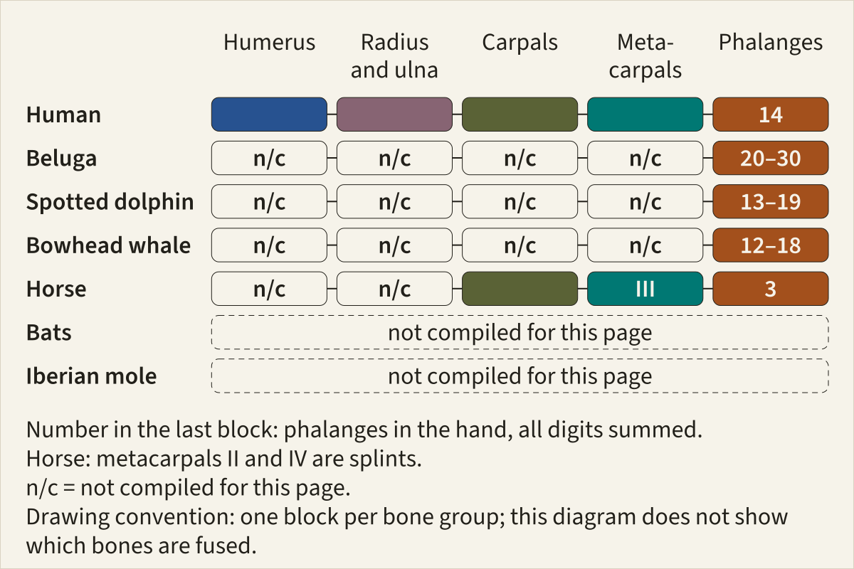
Five forelimbs drawn as chains of bone groups, shoulder to fingertip. A block is coloured only where a source read for this page names that bone group in that animal; the rest are marked n/c. The last block counts the finger bones of the whole hand.
Basis Bone groups and phalanx counts from Gray's Anatomy (1918), de Bakker et al. 2025, Gavazzi et al. 2023, Solounias et al. 2018 and Vincelette et al. 2023; colours fixed site-wide.
Bone colours, site table v1.4humerusradius and ulnacarpalsmetacarpalsphalanges
"Same bones, same colours: five forelimbs" by Feathers, Skeletons & Nests, https://feathersskeletonsandnests.com/skeletons/homologous-structures/ , licensed under CC BY 4.0, https://creativecommons.org/licenses/by/4.0/
A beluga's second finger can hold eight bones in a row. Your index finger holds three. Anatomists still count both as digit II, and that is the core of homologous structures: body parts in different species that trace back to the same part in a shared ancestor, whatever job each one does now.
The standard example is the forelimb of tetrapods, the four-limbed vertebrates. A bat's wing, a whale's flipper, a horse's foreleg and your arm all derive from the same ancestral limb. Each is built in the same order (Solounias et al. 2018): one upper-arm bone (the humerus), two forearm bones (the radius and ulna), a wrist of carpals, a palm of metacarpals, and finger bones called phalanges. The number of phalanges runs from 3 in a horse to between 20 and 30 in an adult beluga.
What did Darwin mean by "the same bones"?
Darwin put the puzzle into a single question in 1859: “What can be more curious than that the hand of a man, formed for grasping, that of a mole for digging, the leg of the horse, the paddle of the porpoise, and the wing of the bat, should all be constructed on the same pattern, and should include the same bones, in the same relative positions?”
Every animal on his list has a row in the table below, with a beluga and a spotted dolphin standing in for his porpoise, although the bat and mole rows are described in words rather than counted. The phrase to hold onto is “relative positions.”
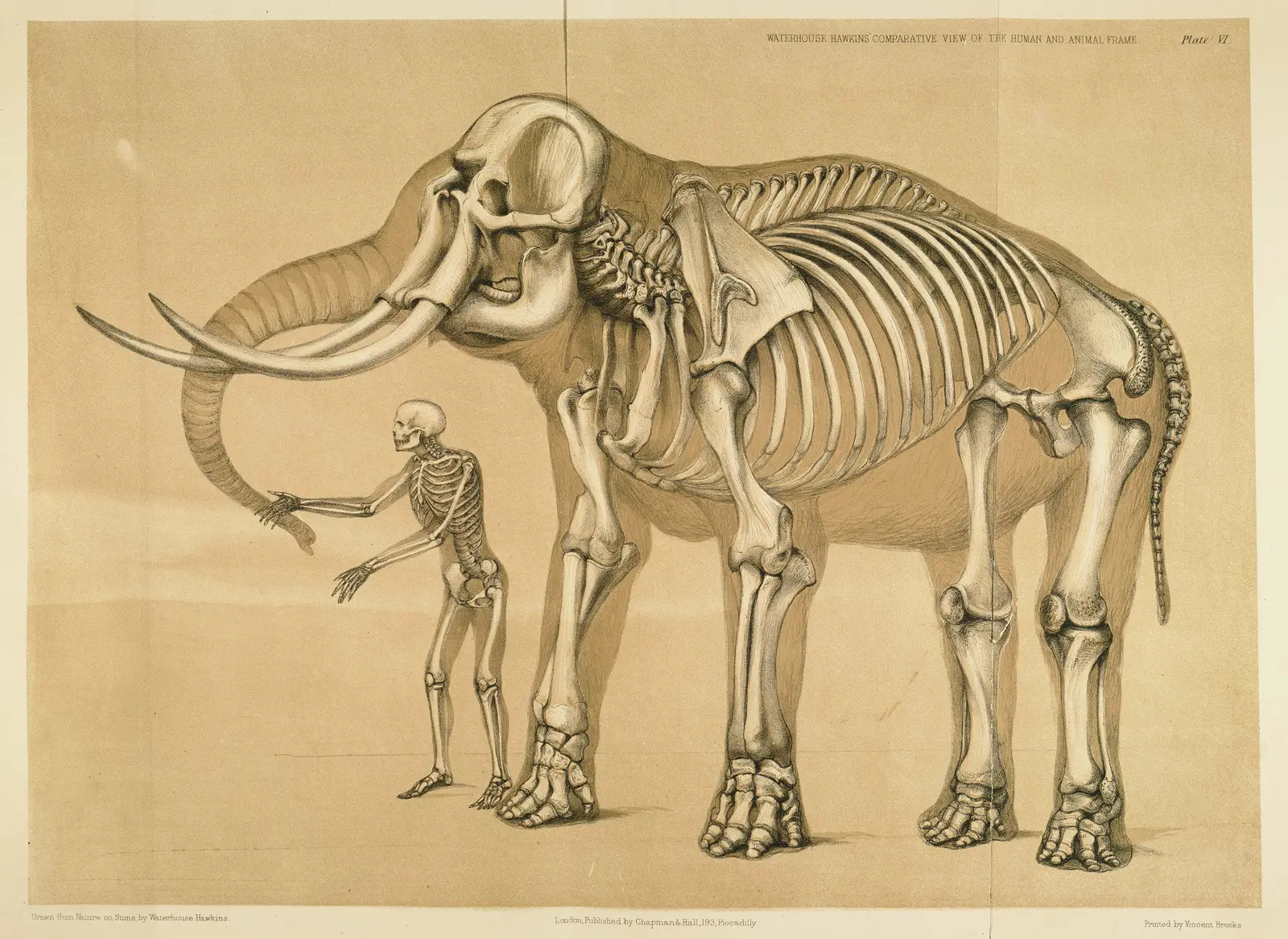
Plate VI of Benjamin Waterhouse Hawkins's Comparative View of the Human and Animal Frame (1860): a human skeleton standing beside an elephant skeleton, the elephant's body drawn as a faint outline around its bones. Follow each front limb down from the shoulder.
"Plate VI, Man and the elephant, from A Comparative View of the Human and Animal Frame" by Benjamin Waterhouse Hawkins — https://commons.wikimedia.org/wiki/File:Comparative_view_of_the_human_and_elephant_frame,_Benjamin_Waterhouse_Hawkins,_1860.jpg — Public domain (US: published 1860; EU: artist Benjamin Waterhouse Hawkins d. 1894, who drew the plate on stone himself (printer Vincent Brooks, dates not checked))
He credited Geoffroy St. Hilaire with the rule behind it. The parts, Darwin wrote, “may change to almost any extent in form and size, and yet they always remain connected together in the same order.” The bones of the arm and forearm, he added, are never found “transposed,” which is why “the same names can be given to the homologous bones in widely different animals.”
The word is older than Darwin's explanation. In his 1843 lectures, Richard Owen defined a homologue as “the same organ in different animals under every variety of form and function,” and an analogue as a part with “the same function” as a part in another animal. Owen's definitions say nothing about ancestry. Darwin's explanation was an ancient progenitor whose limbs were “constructed on the existing general pattern,” then modified by small steps along each line of descent.
Darwin also allowed that the pattern could blur. He named three ways: “the complete abortion of certain parts,” “the soldering together of other parts,” and “the doubling or multiplication of others.” Loss, fusion and multiplication all turn up in the forelimbs below.
Count the finger bones: seven forelimbs and an ancestor
Anatomists record finger bones as a phalangeal formula, the number of phalanges in each digit counted from the thumb side. Your hand is 2-3-3-3-3: two in the thumb and three in each finger, 14 in all (Gray's Anatomy, 1918). Most mammals share that formula (de Bakker et al. 2025). The table adds the inferred formula of the ancestral amniote, the shared ancestor of mammals, reptiles and birds.
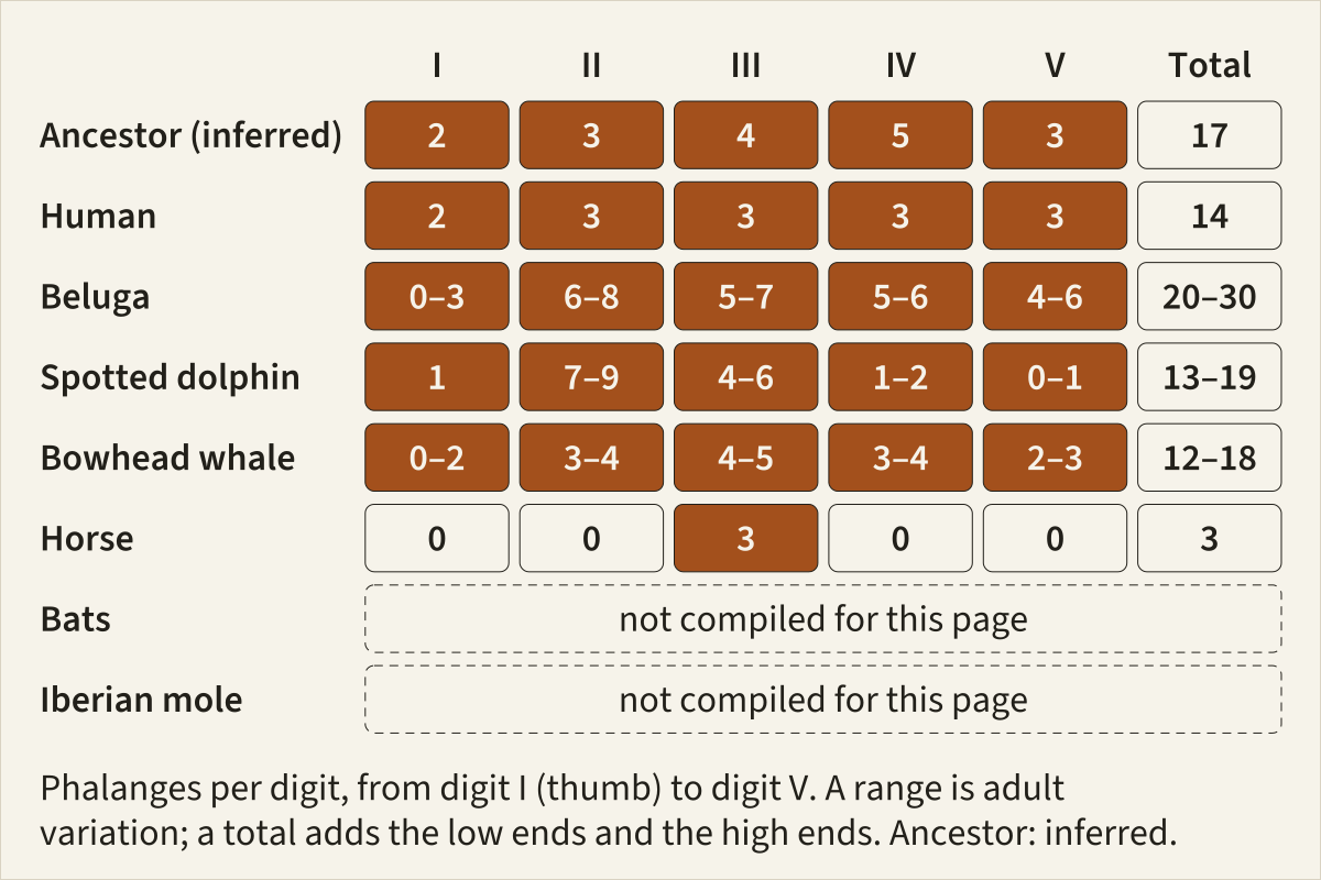
Finger bones per digit, from digit I on the thumb side to digit V, with totals. The rows for bats and the Iberian mole are marked not compiled; the text describes what their studies found.
Basis Counts from Gray's Anatomy (1918), de Bakker et al. 2025, Gavazzi et al. 2023 and Solounias et al. 2018, as listed row by row in the data table; totals recalculated.
"Finger bones per digit in five mammals and an inferred ancestor" by Feathers, Skeletons & Nests, https://feathersskeletonsandnests.com/skeletons/homologous-structures/ , licensed under CC BY 4.0, https://creativecommons.org/licenses/by/4.0/
| Forelimb | I | II | III | IV | V | Total |
|---|---|---|---|---|---|---|
| Ancestral amniote (inferred) | 2 | 3 | 4 | 5 | 3 | 17 |
| Human | 2 | 3 | 3 | 3 | 3 | 14 |
| Beluga, adult | 0–3 | 6–8 | 5–7 | 5–6 | 4–6 | 20–30 |
| Pantropical spotted dolphin, adult | 1 | 7–9 | 4–6 | 1–2 | 0–1 | 13–19 |
| Bowhead whale, adult | 0–2 | 3–4 | 4–5 | 3–4 | 2–3 | 12–18 |
| Horse | 0 | 0 | 3 | 0 | 0 | 3 |
| Bats, 44 species | not compiled | not compiled | not compiled | not compiled | not compiled | not compiled |
| Iberian mole | not compiled | not compiled | not compiled | not compiled | not compiled | not compiled |
Download the phalanx counts (CSV) (what this file records) Data: de Bakker et al. 2025, Mol Biol Evol 42(6): msaf113, CC BY 4.0, https://doi.org/10.1093/molbev/msaf113; Gavazzi et al. 2023, J Morphol 284(2): e21543, CC BY 4.0, https://doi.org/10.1002/jmor.21543; Solounias et al. 2018, R Soc Open Sci 5(1): 171782, CC BY 4.0, https://doi.org/10.1098/rsos.171782; Vincelette et al. 2023, R Soc Open Sci 10(6): 230358, CC BY 4.0, https://doi.org/10.1098/rsos.230358
Each row's counts come from the sources named in the CSV. Totals add the low ends and the high ends of each digit's range, so they are outer limits that no single animal has to reach. The horse's digits II and IV survive as splint bones with no phalanges.
The bat and mole rows stay empty because the studies behind them are paraphrased on this page rather than tabulated. The sections below say what they found. The sources read for this table give no mole-specific count.
Why does a beluga have more finger bones than you?
A beluga's flipper is its forelimb. In cetaceans, the whales, dolphins and porpoises, a soft-tissue flipper encases most of the forelimb (Cooper et al. 2007), and that flipper evolved from a five-fingered, weight-bearing limb in early amphibious whales (Gavazzi et al. 2023).
The extra bones have a name: hyperphalangy, an increase in the number of phalanges over the standard mammal formula of 2-3-3-3-3 (Gavazzi et al. 2023). Telizhenko and colleagues (2024), citing earlier work, note that among mammals extra phalanges turn up only in cetaceans. Elsewhere, they add, changes to the mammal formula mostly remove bones, as in bats and hoofed mammals.
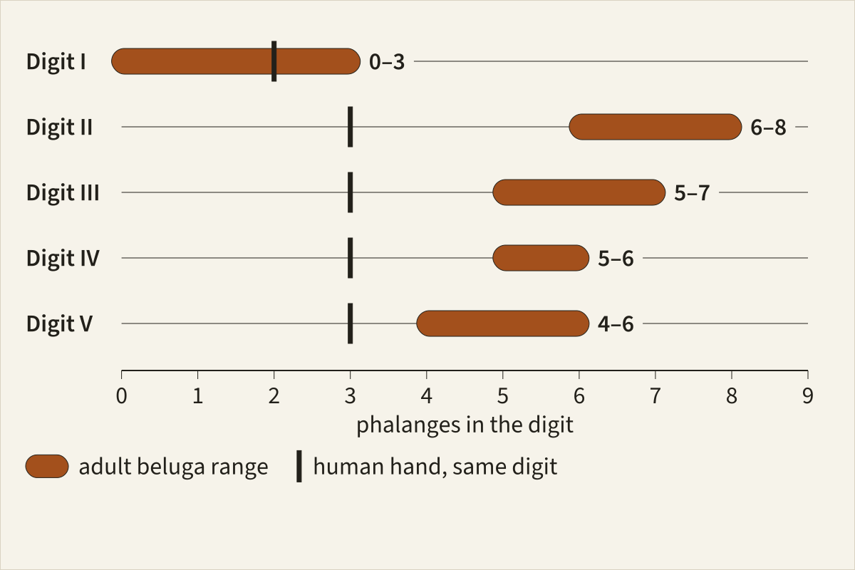
Adult beluga ranges of finger bones in each digit, with the count for the same digit of a human hand marked as a short bar.
Basis Beluga ranges as compiled by Gavazzi et al. 2023; human counts from Gray's Anatomy (1918).
"Beluga finger bones per digit beside the human count" by Feathers, Skeletons & Nests, https://feathersskeletonsandnests.com/skeletons/homologous-structures/ , licensed under CC BY 4.0, https://creativecommons.org/licenses/by/4.0/
The beluga's adult range runs 0–3, 6–8, 5–7, 5–6 and 4–6 from digit I to digit V, figures compiled from older anatomical studies by Gavazzi and colleagues (2023). Digit II is both its longest digit and the one with the most phalanges. Put that next to your index finger: three bones in yours, six to eight in the beluga's.
A flipper does not only add. The pantropical spotted dolphin carries 7–9 phalanges in digit II but only 1–2 in digit IV and 0–1 in digit V, fewer than your ring and little fingers. The bowhead whale, a baleen whale, consistently adds bones in digit III, and its summed range of 12 to 18 straddles your 14.
In spotted dolphin embryos, Richardson and Oelschläger (2002) confirmed earlier reports that phalanges keep being added for longer in digits II and III. They also found that new phalanges form only at the tips.
Across cetaceans, Cooper and colleagues (2007) found the most extra phalanges in digits II and III of toothed whales and in digits III and IV of baleen whales. Most baleen whales have four digits, having lost every element of digit I, its metacarpal included.
Do bat wings have extra finger bones?
A bat's wing is a membrane stretched between long fingers, and digits III, IV and V support most of it (Bahlman et al. 2016). Fingers that long might seem to need extra bones. They have fewer.
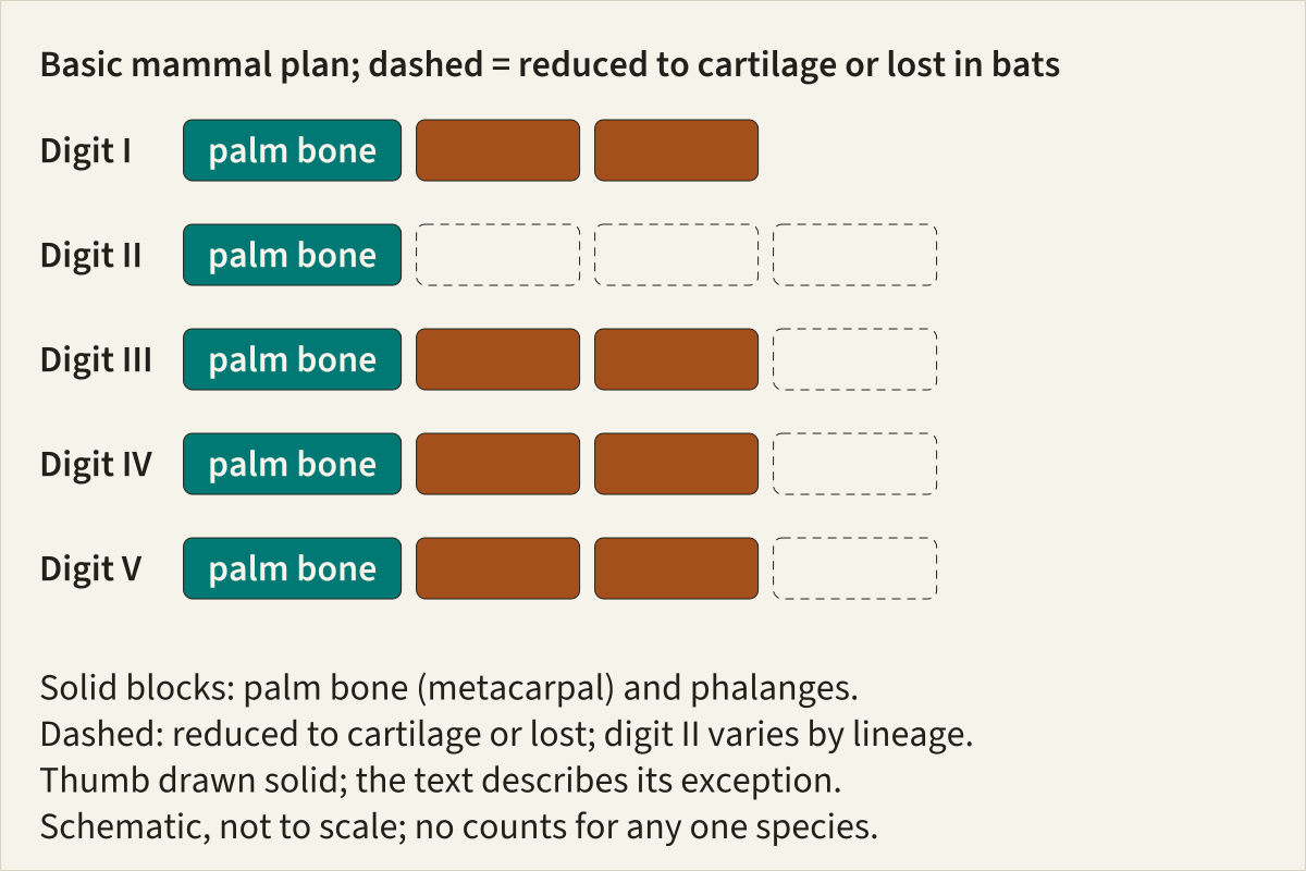
The basic mammal hand drawn as blocks, with dashed outlines on the bones that bat lineages have reduced to cartilage or lost; on digit II, how many depends on the lineage. The thumb is drawn solid; the text describes its exception. Positions only; no counts for any single species.
Basis Basic mammal formula from de Bakker et al. 2025; reduced bones placed after the description in Bahlman et al. 2016; schematic, not to scale.
"Where bat wings lose finger bones: a schematic" by Feathers, Skeletons & Nests, https://feathersskeletonsandnests.com/skeletons/homologous-structures/ , licensed under CC BY 4.0, https://creativecommons.org/licenses/by/4.0/
Bahlman and colleagues dissected the wings of 44 bat species from 17 of the 20 living bat families. Every species had lost at least one finger joint, along with its phalanx, compared with the basic mammal hand. Digit II varies most, having lost one to three of its phalanges depending on the lineage.
On digits III to V, only the third and outermost phalanx was ever lost. In about a third of the species sampled it was gone entirely, and in the rest it had shrunk to a flexible tip of cartilage. Only one species in the survey, Amorphochilus schnablii of the family Furipteridae, had lost the phalanges of its thumb.
Bats show Darwin's “soldering” too. In many species the last phalanx is reduced to a small piece of cartilage fused to the end of the bone before it, as earlier anatomists described and Bahlman and colleagues summarise.
How does a horse stand on one finger?
A horse walks on digit III alone. That digit has three phalanges, which horse anatomy calls the long pastern, the short pastern and the coffin bone inside the hoof (Solounias et al. 2018). Beside its main palm bone, metacarpal III or the cannon bone, sit two thin splint bones: what remains of the metacarpals of digits II and IV (Vincelette et al. 2023).
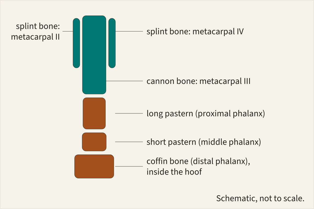
The bones below a horse's wrist: the cannon bone with a splint bone on each side, then the long pastern, the short pastern and the coffin bone.
Basis Bone names from Solounias et al. 2018 and Vincelette et al. 2023; schematic, not to scale.
"A horse's single digit: cannon bone, splints and three phalanges" by Feathers, Skeletons & Nests, https://feathersskeletonsandnests.com/skeletons/homologous-structures/ , licensed under CC BY 4.0, https://creativecommons.org/licenses/by/4.0/
That is the standard reading. The 3 in the table follows it. A 2018 study proposed that parts of all five digits survive, reading the frog (a V-shaped structure on the underside of the hoof) as digits II and IV, and the wings and cartilages of the coffin bone as digits I and V.
A 2023 response accepted that upper remnants of all four side digits survive, digits I and V as ridges on the splint bones, but found no evidence for the hoof reading, noting that the hoof wall, sole and frog form much earlier in development than the fetal stages the 2018 team examined (Vincelette et al. 2023). Either way, the dispute concerns the hoof, and neither reading adds a separate finger bone to the count.
Here the old-house picture helps, as long as you read it as a picture. Think of a forelimb as a house renovated room by room while people still live in it: walls come down and boards go up, yet the floor plan stays. The horse's splints are rooms that were closed off and never torn out. In anatomical terms, the side metapodials kept their upper parts and lost their phalanges, and they still help support the wrist (Vincelette et al. 2023).
Bones that shrink but keep a job have their own page, on vestigial structures.
Does a mole have a sixth finger?
A mole's hand looks as if it has a sixth digit. Moles in several genera of the family Talpidae carry a large, sickle-shaped bone on the thumb side of the hand, the radial sesamoid, also called the os falciforme (Mitgutsch et al. 2012). Gray's Anatomy (1918, p. 277) defines sesamoid bones as “small more or less rounded masses embedded in certain tendons and usually related to joint surfaces,” and names the patella as the largest sesamoid bone of the lower limb.
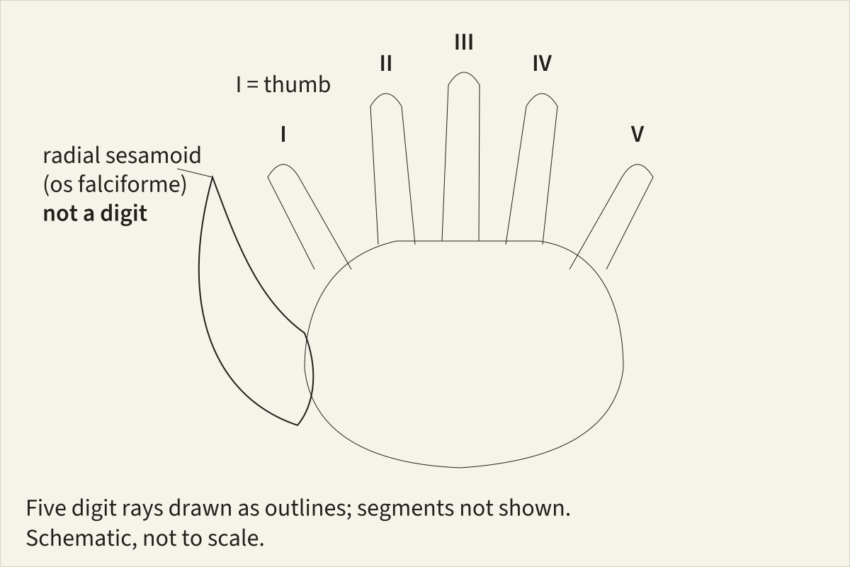
A mole's hand with its five digit rays and, beside the thumb, the sickle-shaped radial sesamoid. Outlines only.
Basis Position of the radial sesamoid after the description in Mitgutsch et al. 2012, drawn independently; schematic, not to scale.
"A mole's hand: five digits and a radial sesamoid" by Feathers, Skeletons & Nests, https://feathersskeletonsandnests.com/skeletons/homologous-structures/ , licensed under CC BY 4.0, https://creativecommons.org/licenses/by/4.0/
The mole's version widens the hand and braces the animal as it digs, and in the most specialised moles it can be moved on its own like a digit. It still lacks the segments of a finger, and in embryos of the Iberian mole it begins forming later than the true digits. The hand still has five fingers; in these moles a wrist sesamoid was drawn into the row of digits.
The remodel rule is a first question to put to any odd bone: what existing structure was it remodelled from? Asked of the mole, it points to a wrist sesamoid instead of a new finger. It is a habit for reading skeletons, not a law of evolution, so it can still point the wrong way.
Some remodelled bones go further and change jobs outright. A bone that took on a new job in a lineage and stayed in the body to do it is what this site calls a stowaway bone; the malleus and incus of the mammal middle ear, once part of the jaw joint, are the example on the middle-ear page linked below.
Your hand is a trimmed version too
Your own 2-3-3-3-3 is not the starting point. The ancestral amniote probably had a 2-3-4-5-3 hand (de Bakker et al. 2025). Compared with that, the typical mammal hand has lost one phalanx from digit III and two from digit IV.
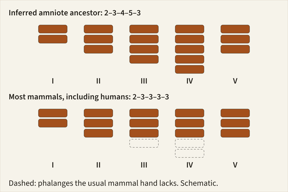
The inferred ancestral amniote hand, 2-3-4-5-3, above the usual mammal hand, 2-3-3-3-3; dashed blocks mark the phalanges missing from digits III and IV.
Basis Formulas from de Bakker et al. 2025; schematic, not to scale.
"From the inferred amniote hand to the usual mammal hand" by Feathers, Skeletons & Nests, https://feathersskeletonsandnests.com/skeletons/homologous-structures/ , licensed under CC BY 4.0, https://creativecommons.org/licenses/by/4.0/
Your hand holds 27 bones in all, not counting the small sesamoids that Gray's Anatomy (1918) lists separately: 8 carpals, 5 metacarpals and 14 phalanges. Line the totals up and your 14 sits mid-table. It is above the horse, and above every bat in the survey, since each species there had lost at least one phalanx (Bahlman et al. 2016). It is below the beluga and the inferred ancestor.
How do biologists decide two bones are the same bone?
Counting cannot settle homology, because counts change. From the early 1800s into the first half of the 1900s, anatomists relied on two main tests: position, meaning a bone keeps the same neighbours, and development, meaning it grows from the same embryonic precursor (Brigandt 2011). In a limb, position carries the argument Darwin took from Geoffroy: a bone that sits between the wrist and a finger is a metacarpal, however long or short it has become.

Owen's 1843 glossary, page 379: the entry for Homologue, between Homogangliate and Homomorphous.
"Glossary, p. 379 (entry Homologue), from Lectures on the Comparative Anatomy and Physiology of the Invertebrate Animals" by Richard Owen — https://archive.org/details/lecturesoncompar00owen/page/n386/ — Public domain (US: published 1843; EU: author Richard Owen d. 1892; Internet Archive metadata marks the item NOT_IN_COPYRIGHT); modified: cropped to the Homologue entry and the entries on either side
Owen's two glossary entries also show how a bird's wing and a bat's wing can be both at once. Both are forelimbs built on the tetrapod sequence of humerus, radius and ulna, wrist and hand bones, which Solounias and colleagues describe for tetrapod forelimbs in general, so as forelimbs they are homologues. Both are used to fly, so as wings they are analogues: parts with the same function. The bird's version is traced bone by bone on bird wing bones.
The forelimb sequence on this page is a tetrapod sequence. How it relates to the bones inside a fish fin is a question for fossils such as Tiktaalik.
Same bones, elsewhere:
- Middle ear evolution: the quadrate and articular of early synapsids match the incus and malleus in your middle ear, two stowaway bones.
- Are birds dinosaurs?: where bats, whales and horses branch off is a family-tree question, and on that kind of tree birds sit inside the dinosaurs.
Sources
- Charles Darwin, On the Origin of Species by Means of Natural Selection, 1st edition (London: John Murray, 1859), chapter XIII. gutenberg.pglaf.org/cache/epub/1228/pg1228.txtRead: full text of chapter XIII (sections Morphology and Rudimentary, atrophied, or aborted organs)
- Richard Owen, Lectures on the Comparative Anatomy and Physiology of the Invertebrate Animals, Delivered at the Royal College of Surgeons, in 1843 (London: Longman, Brown, Green, and Longmans, 1843), glossary entries "Analogue" (p. 374) and "Homologue" (p. 379). archive.org/details/lecturesoncompar00owenRead: pages 374 and 379 (glossary), checked against the page images
- Nikos Solounias, Melinda Danowitz, Elizabeth Stachtiaris, Abhilasha Khurana, Marwan Araim, Marc Sayegh and Jessica Natale, "The evolution and anatomy of the horse manus with an emphasis on digit reduction", Royal Society Open Science 5, no. 1 (2018): 171782. doi.org/10.1098/rsos.171782Read: full textText licence: CC BY 4.0 (terms)
- Henry Gray, Anatomy of the Human Body, 20th edition, edited by Warren H. Lewis (Philadelphia: Lea & Febiger, 1918), pp. 219, 221, 227, 230, 266–267, 277 and 1044. archive.org/details/anatomyofhumanbo1918grayRead: pages 219, 221, 227, 230, 266, 267, 277 and 1044, checked against the page images
- Merijn A. G. de Bakker, Luthfi Nurhidayat, Alisha Kiran Dijkerman, Wing Yu Chung, Elena C. Oudesluys, Kaylah de Jager, Joost Willemse and Michael K. Richardson, "Changes in evolutionary developmental control points in the amniote limb may explain hyperphalangy", Molecular Biology and Evolution 42, no. 6 (2025): msaf113. doi.org/10.1093/molbev/msaf113Read: full textText licence: CC BY 4.0 (terms)
- Lia Gavazzi, Lisa N. Cooper, Sharon Usip, Robert Suydam, Raphaela Stimmelmayr, John C. George, Greg O'Corry-Crowe, Chih-Wei Hsu and Johannes Thewissen, "Comparative embryology of Delphinapterus leucas (beluga whale), Balaena mysticetus (bowhead whale), and Stenella attenuata (pan-tropical spotted dolphin) (Cetacea: Mammalia)", Journal of Morphology 284, no. 2 (2023): e21543. doi.org/10.1002/jmor.21543Read: full textText licence: CC BY 4.0 (terms)
- Lisa Noelle Cooper, Annalisa Berta, Susan D. Dawson and Joy S. Reidenberg, "Evolution of hyperphalangy and digit reduction in the cetacean manus", The Anatomical Record 290, no. 6 (2007): 654–672. doi.org/10.1002/ar.20532Read: abstract only
- Valeriia Telizhenko, Carolin Kosiol, Michael R. McGowen and Pavel Gol'din, "Relaxed selection in evolution of genes regulating limb development gives clue to variation in forelimb morphology of cetaceans and other mammals", Proceedings of the Royal Society B 291, no. 2032 (2024): 20241106. doi.org/10.1098/rspb.2024.1106Read: full text
- Michael K. Richardson and Helmut H. A. Oelschläger, "Time, pattern, and heterochrony: a study of hyperphalangy in the dolphin embryo flipper", Evolution & Development 4, no. 6 (2002): 435–444. doi.org/10.1046/j.1525-142x.2002.02032.xRead: abstract only
- Joseph W. Bahlman, Rosalyn M. Price-Waldman, Hannah W. Lippe, Kenneth S. Breuer and Sharon M. Swartz, "Simplifying a wing: diversity and functional consequences of digital joint reduction in bat wings", Journal of Anatomy 229, no. 1 (2016): 114–127. doi.org/10.1111/joa.12457Read: full text
- Alan R. Vincelette, Elise Renders, Kathleen M. Scott, Peter L. Falkingham and Christine M. Janis, "Hipparion tracks and horses' toes: the evolution of the equid single hoof", Royal Society Open Science 10, no. 6 (2023): 230358. doi.org/10.1098/rsos.230358Read: full textText licence: CC BY 4.0 (terms)
- Christian Mitgutsch, Michael K. Richardson, Rafael Jiménez, José E. Martin, Peter Kondrashov, Merijn A. G. de Bakker and Marcelo R. Sánchez-Villagra, "Circumventing the polydactyly 'constraint': the mole's 'thumb'", Biology Letters 8, no. 1 (2012): 74–77. doi.org/10.1098/rsbl.2011.0494Read: full text
- Ingo Brigandt, "Essay: Homology", Embryo Project Encyclopedia (Arizona State University, 2011). embryo.asu.edu/pages/essay-homologyRead: bibliography only
- Aitor Navarro-Díaz, Borja Esteve-Altava and Diego Rasskin-Gutman, "Disconnecting bones within the jaw-otic network modules underlies mammalian middle ear evolution", Journal of Anatomy 235, no. 1 (2019): 15–33. doi.org/10.1111/joa.12992Read: full text
Cite this page
Feathers, Skeletons & Nests. “Homologous Structures: Same Bones, Different Counts.” first published 17 November 2026. https://feathersskeletonsandnests.com/skeletons/homologous-structures/ (accessed on the date you read it).
How facts are sourcedAll correctionsReport an error on this page (contact@feathersskeletonsandnests.com)
More from the Skeletons drawer
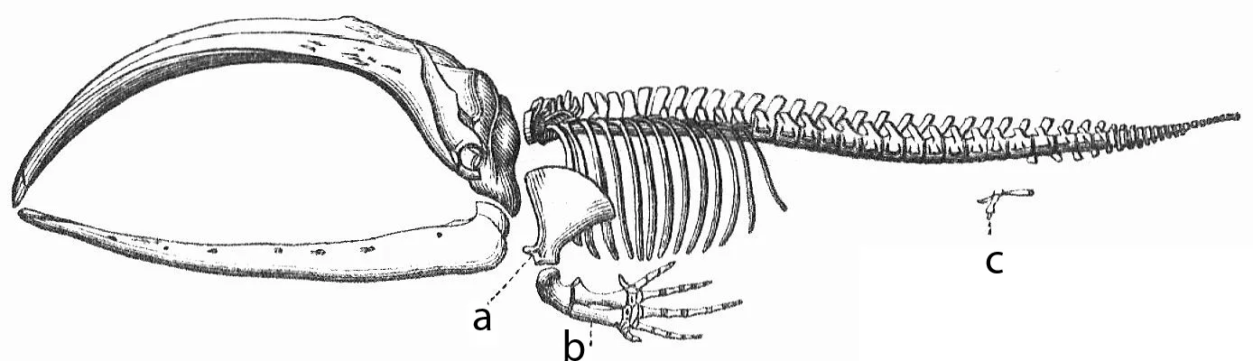
Vestigial Structures: A Whale’s Hip Bones Anchor Muscles
- Kind
- Comparison and reading
- Added
Picture credit
"Skeleton of a baleen whale, from Meyers Konversations-Lexikon, 4th edition" by Unknown author, Meyers Konversations-Lexikon (1888) — https://commons.wikimedia.org/wiki/File:Whale_skeleton.png — Public domain (US: published 1888, before 1931; EU: anonymous work published 1888, more than 70 years ago)
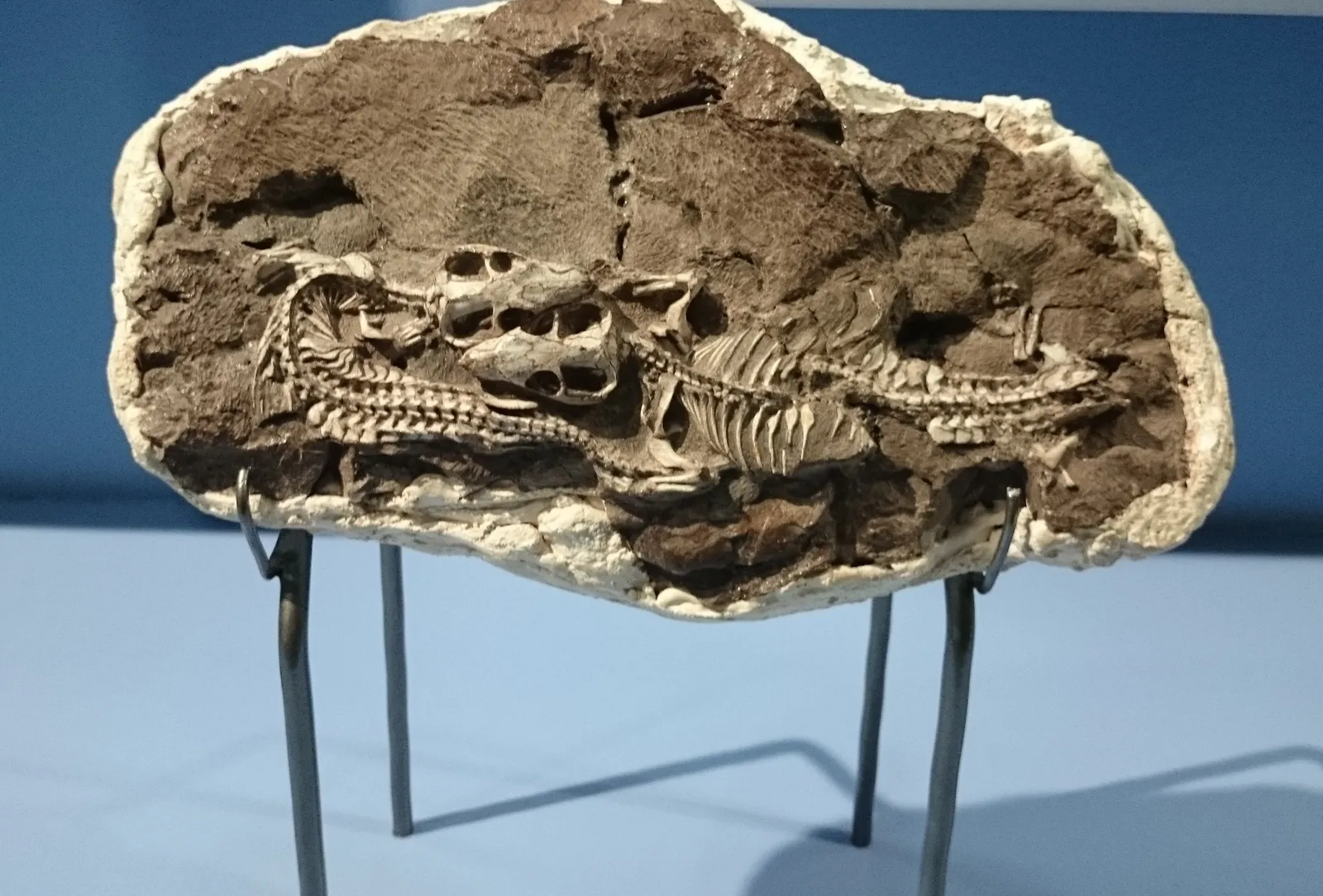
Middle Ear Evolution: Two Jaw Bones Became Ear Bones
- Kind
- In-depth explainer
- Added
Picture credit
"Thrinaxodon fossil, Iziko South African Museum" by Nkansahrexford — https://commons.wikimedia.org/wiki/File:Iziko_Thrinaxodon_fossil.JPG — CC BY 3.0 (https://creativecommons.org/licenses/by/3.0/); modified: cropped: top 120 px removed (museum display panel with printed text above the fossil block)
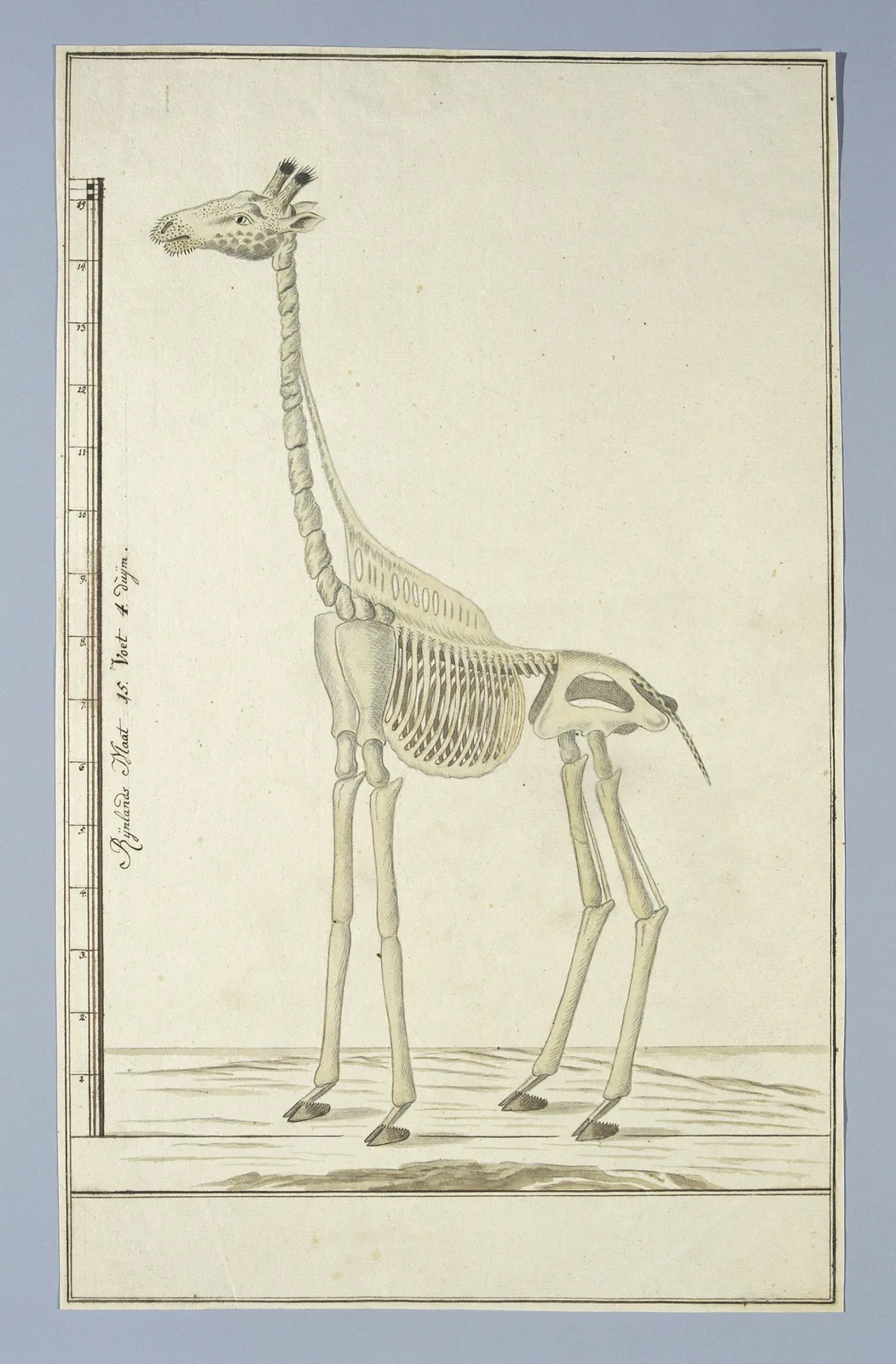
How Many Bones in a Giraffe Neck? Seven, Like Most Mammals
- Kind
- Specimen entry
- Added
Picture credit
"Giraffa camelopardalis (Giraf), skelet, from the Robert Jacob Gordon drawings" by Robert Jacob Gordon / Rijksmuseum — https://commons.wikimedia.org/wiki/File:Giraffa_camelopardalis_(Giraf),_skelet,_RP-T-1914-17-152.jpg — CC0 1.0 (https://creativecommons.org/publicdomain/zero/1.0/)