Middle Ear Evolution: Two Jaw Bones Became Ear Bones
Short answer
In the lineage that led to mammals, the articular and quadrate of the old jaw joint became the middle ear's malleus and incus, as a 2017 review by Abigail Tucker sets out. The third ear bone, the stapes, came from elsewhere. Early relatives such as Morganucodon had a double jaw joint, old and new hinges side by side; this is an ancestral line, not living reptiles turning into mammals.
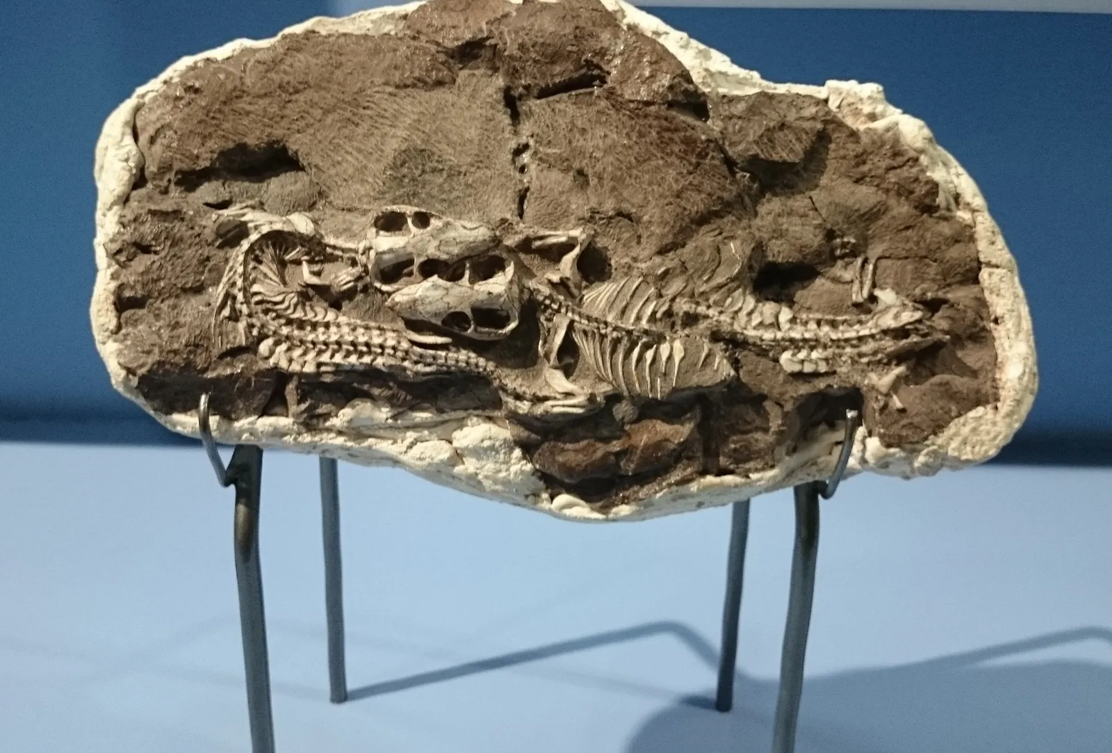
A block of rock holding Thrinaxodon fossils, on display at the Iziko South African Museum in Cape Town. Two small skulls sit near the middle of the block, with backbones and ribs curling around them.
"Thrinaxodon fossil, Iziko South African Museum" by Nkansahrexford — https://commons.wikimedia.org/wiki/File:Iziko_Thrinaxodon_fossil.JPG — CC BY 3.0 (https://creativecommons.org/licenses/by/3.0/); modified: cropped: top 120 px removed (museum display panel with printed text above the fossil block)
Two of the three bones in your middle ear started out as parts of a jaw joint. They are the malleus and the incus. The 1918 edition of Gray's Anatomy traces their names to a supposed likeness to a hammer and an anvil, and lists them with the stapes as a chain of three movable bones. In earlier relatives of mammals, the matching bones were the articular, at the back of the lower jaw, and the quadrate, an upper-jaw bone that the articular hinged against.
That history runs through long-extinct ancestors. Living reptiles sit on a separate branch of the amniotes, the group that also holds birds and mammals, so setting a lizard's jaw beside your ear compares matching bones and does not show one turning into the other. Thrinaxodon, the animal in the photo, is one of 43 synapsids whose jaw and ear bones a 2019 study by Aitor Navarro-Díaz and colleagues mapped. Synapsids are the group that includes mammals and their extinct relatives.
Which ear bones came from the jaw?
The malleus and the incus did. A 2017 review of middle-ear evolution by Abigail Tucker of King's College London states that the articular and the quadrate are homologous to the malleus and the incus: they are the same bones, inherited from a shared ancestor, like other homologous structures. Reichert first proposed that match and Gaupp extended it, as summarised by Tucker 2017.
Other jaw bones moved into supporting roles. In reptiles, the bone called the angular matches the tympanic ring, the bony frame that holds the mammal eardrum. The prearticular matches the gonial, a bone associated with that ring. A third jaw bone, the surangular, is thought to match the accessory malleus, a small extra bone above the malleus found in a few mammals but not in the mouse.
All of those jobs once belonged to the lower jaw. In mammals a single bone, the dentary, does the work that the angular, surangular, splenial and prearticular shared in other jawed vertebrates, Tucker writes. Bones from behind the dentary, such as the angular and the prearticular, ended up with new roles in the ear.
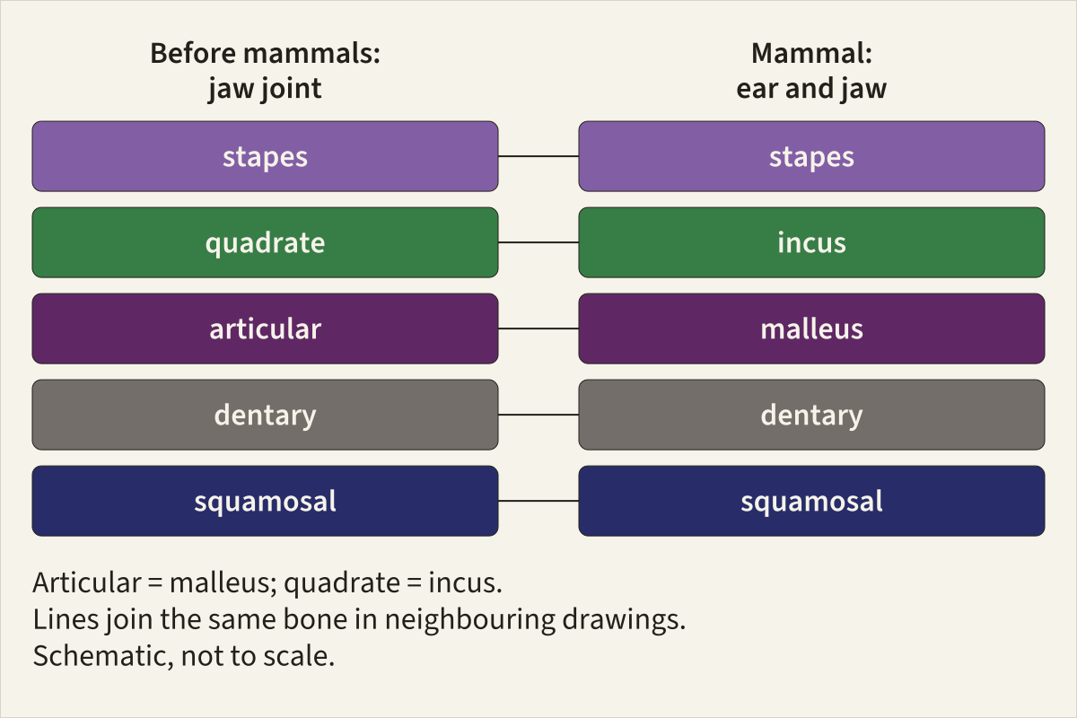
Five bones in two states: the jaw joint before mammals and the mammal ear and jaw. Each row joins one bone in both states, so the quadrate row ends at the incus and the articular row ends at the malleus.
Basis Bones and their homologues as summarised by Tucker 2017; schematic, not to scale; colours fixed site-wide.
Bone colours, site table v1.4dentaryarticular, the same bone as the malleusquadrate, the same bone as the incussquamosalstapes
"Jaw joint and middle ear: the same bones in two states" by Feathers, Skeletons & Nests, https://feathersskeletonsandnests.com/skeletons/middle-ear-evolution/ , licensed under CC BY 4.0, https://creativecommons.org/licenses/by/4.0/
Data: Tucker A. S. (2017) Major evolutionary transitions and innovations: the tympanic middle ear. Philosophical Transactions of the Royal Society B 372: 20150483, https://doi.org/10.1098/rstb.2015.0483, CC BY 4.0
| Bone before mammals | Bone in mammals | Evidence | Source |
|---|---|---|---|
| Articular | Malleus | Comparative anatomy; malleus and incus form on Meckel's cartilage with a joint between them, as articular and quadrate do; shared joint genes | Tucker 2017, section 7 |
| Quadrate | Incus | Comparative anatomy; malleus and incus form on Meckel's cartilage with a joint between them, as articular and quadrate do; shared joint genes | Tucker 2017, section 7 |
| Angular | Tympanic ring (ectotympanic) | How the bones form, their position, timing and gene expression | Tucker 2017, section 2 |
| Prearticular | Gonial | How the bones form, their position, timing and gene expression | Tucker 2017, section 2 |
| Surangular | Accessory malleus (a few mammals) | Proposed match only | Tucker 2017, section 9 |
| Stapes (reptiles), columella (birds) | Stapes | Same ear bone, not from the jaw joint; mostly second pharyngeal arch | Tucker 2017, section 1 |
Download the jaw-to-ear homologies (CSV) (what this file records) Data: Tucker A. S. (2017) Major evolutionary transitions and innovations: the tympanic middle ear. Philosophical Transactions of the Royal Society B 372: 20150483, https://doi.org/10.1098/rstb.2015.0483, CC BY 4.0
What is a stowaway bone?
Feathers, Skeletons & Nests calls a bone like the malleus or the incus a stowaway bone, a bone that took on a new job in a lineage and stayed in the body to do it. Think of an old beam taken down during a renovation and carried into another room, where it now holds up something else. The comparison describes the result only. It says nothing about how the change happened.
The old hinge came along with the bones. In the embryo, Tucker writes, the malleus and the incus form with a joint between them in the same way as the articular and the quadrate do in other jawed vertebrates. The small joint between your malleus and incus therefore corresponds to the jaw hinge of earlier relatives. It no longer opens and closes a mouth.
The stapes is left out of this case on purpose. It has a different origin, set out in its own section below. Ribs that took on a new job as a shell are another possible candidate for the term, covered in the entry on the turtle shell.
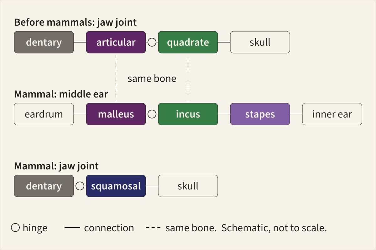
The two states drawn as connections and hinges. In the top row the hinge sits between the articular and the quadrate. In the middle row the same two bones, now the malleus and the incus, keep a joint between them inside the ear, and in the bottom row the mammal jaw swings on a new hinge.
Basis Bones, joints and homologues as summarised by Tucker 2017; schematic, not to scale; colours fixed site-wide.
Bone colours, site table v1.4dentaryarticular, the same bone as the malleusquadrate, the same bone as the incussquamosalstapes
"The jaw hinge before mammals and the same two bones inside the mammal ear" by Feathers, Skeletons & Nests, https://feathersskeletonsandnests.com/skeletons/middle-ear-evolution/ , licensed under CC BY 4.0, https://creativecommons.org/licenses/by/4.0/
Data: Tucker A. S. (2017) Major evolutionary transitions and innovations: the tympanic middle ear. Philosophical Transactions of the Royal Society B 372: 20150483, https://doi.org/10.1098/rstb.2015.0483, CC BY 4.0
How could an animal chew with ear bones in its jaw?
It used two hinges at once. Tucker writes that basal mammaliforms, such as Morganucodon, show a double jaw joint in the fossil record. One hinge was the old joint between the articular and the quadrate. The other was a new joint between an enlarged dentary and the squamosal, a bone of the upper jaw.
Mammals can chew by moving the lower jaw sideways as well as up and down, and their upper and lower teeth often fit together for grinding. Toothed non-mammals, Tucker notes, generally move the jaw only up and down. In the review's account, the new joint appears to have steadied the jaw, allowing more movement and helping to prevent dislocation. Shearing contact between complex teeth behind the canines appears only after the second joint.
In humans, that new joint is the temporomandibular joint. It is proposed, Tucker writes, that the new joint freed the old one to start playing a role in hearing.
Other fossils show possible steps toward the second hinge. Tucker points to Ictidopsis, a non-mammalian synapsid whose dentary extends upward and might represent a step toward the new joint. She also names two groups, the tritheledontids and brasilodontids, with a ridge on the dentary that touches the squamosal and forms a hinge-like structure. The 2019 study by Navarro-Díaz and colleagues notes that the whole series of changes from jaw to ear started about 315 million years ago.
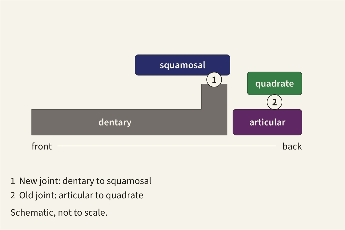
A double jaw joint drawn from the side, front to the left. Hinge 1 is the new joint between the dentary and the squamosal; hinge 2, just behind it, is the old joint between the articular and the quadrate.
Basis Joints as described for Morganucodon-type jaws by Tucker 2017; schematic, not to scale; colours fixed site-wide.
Bone colours, site table v1.4dentaryarticular, the same bone as the malleusquadrate, the same bone as the incussquamosal
"A double jaw joint: a new dentary-squamosal hinge beside the old articular-quadrate hinge" by Feathers, Skeletons & Nests, https://feathersskeletonsandnests.com/skeletons/middle-ear-evolution/ , licensed under CC BY 4.0, https://creativecommons.org/licenses/by/4.0/
Data: Tucker A. S. (2017) Major evolutionary transitions and innovations: the tympanic middle ear. Philosophical Transactions of the Royal Society B 372: 20150483, https://doi.org/10.1098/rstb.2015.0483, CC BY 4.0
How jaw fossils like these were preserved in rock is the subject of how fossils form.
What do embryos show?
Embryos repeat the old arrangement before they leave it. In non-mammalian amniotes, the reptiles and birds, the articular and quadrate form at the back end of Meckel's cartilage, a rod of cartilage in the developing lower jaw. The articular stays attached to that rod for the animal's whole life, Tucker writes.
Mammal embryos start the same way. The malleus and the incus develop connected to Meckel's cartilage, with a joint forming between them. A key joint gene, Bapx1, is expressed both in the jaw joint of non-mammals and in the ear joint of mammals. The 1918 edition of Gray's Anatomy records the human version: in the fetus, the anterior process is the longest process of the malleus and runs straight on into Meckel's cartilage (p. 1044).
Then the mammal ear bones let go. In the mouse, the part of Meckel's cartilage next to the ear bones turns into a ligament, the sphenomandibular ligament, soon after birth. Marsupials are born before the new jaw joint forms, so a newborn's jaw is centred on the ear bones while they are still cartilage. Tucker adds that these probably do not work as a true jaw joint.
The switch can be timed in the opossum Monodelphis. Its middle ear has filled with air by 26 days after birth, and the young can hear by 28 to 30 days, which shows the bones have moved from a jaw job to an ear job.
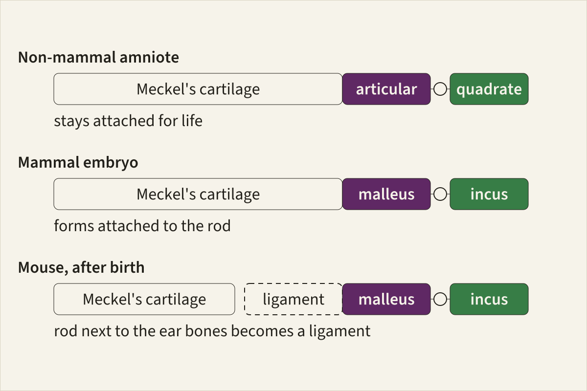
Meckel's cartilage in three settings. In a non-mammal amniote the articular stays on the rod for life; in a mammal embryo the malleus forms on the same rod; in a mouse after birth the stretch of rod next to the ear bones becomes a ligament.
Basis Development as summarised by Tucker 2017; schematic, not to scale.
"Meckel's cartilage and the two jaw-joint elements at its back end" by Feathers, Skeletons & Nests, https://feathersskeletonsandnests.com/skeletons/middle-ear-evolution/ , licensed under CC BY 4.0, https://creativecommons.org/licenses/by/4.0/
Data: Tucker A. S. (2017) Major evolutionary transitions and innovations: the tympanic middle ear. Philosophical Transactions of the Royal Society B 372: 20150483, https://doi.org/10.1098/rstb.2015.0483, CC BY 4.0
Where did the stapes come from?
The stapes was not one of the jaw-joint bones, which is why this page keeps it apart from the two stowaway bones. Reptiles and birds have a single middle-ear bone, called the stapes in reptiles and the columella in birds, and Tucker states that the mammal stapes is homologous to it.
The embryo shows the difference. Cell-grafting experiments in the chick, which follow cells from where they start to where they end up, trace the columella to the second pharyngeal arch, the embryonic arch just behind the one that builds the jaw. Its base comes from mesoderm instead. Genetic labelling in the mouse gives the same pattern for the stapes, while the malleus and incus come largely from the first arch, Tucker writes.
Before hearing, the stapes seems to have had another job. In fossils of early stem amniotes, Tucker writes, a robust, large stapes derived from the hyomandibular shows no sign of use for airborne sound. Instead it appears to have helped steady the skull during biting, and as skulls changed, it was freed from that role and linked to the inner ear.
Reptiles do not share the full set of mammal ear bones. They have a stapes, but no malleus or incus: their articular and quadrate are still jaw bones, and in reptiles and birds that have an eardrum, the quadrate also supports it. Tucker adds that ears with an eardrum arose separately in the different amniote groups during the Early Triassic, so a lizard's ear is not an early draft of yours.
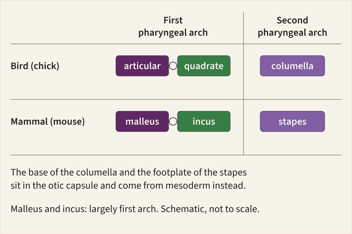
Where each bone mostly starts in the embryo. The articular and quadrate of the chick and the malleus and incus of the mouse sit in the first-arch column; the columella and the stapes sit in the second, apart from a base that comes from mesoderm.
Basis Arch origins in the chick and the mouse as summarised by Tucker 2017; schematic, not to scale; colours fixed site-wide.
Bone colours, site table v1.4articular, the same bone as the malleusquadrate, the same bone as the incusstapes
"Where the ear and jaw-joint bones come from in the embryo, by pharyngeal arch" by Feathers, Skeletons & Nests, https://feathersskeletonsandnests.com/skeletons/middle-ear-evolution/ , licensed under CC BY 4.0, https://creativecommons.org/licenses/by/4.0/
Data: Tucker A. S. (2017) Major evolutionary transitions and innovations: the tympanic middle ear. Philosophical Transactions of the Royal Society B 372: 20150483, https://doi.org/10.1098/rstb.2015.0483, CC BY 4.0
| Animal | First pharyngeal arch | Second pharyngeal arch | Mesoderm (part sitting in the otic capsule) |
|---|---|---|---|
| Bird (chick) | Articular, quadrate | Columella | Base of the columella |
| Mammal (mouse) | Malleus, incus (largely) | Stapes (largely) | Footplate of the stapes |
Who worked this out, and how?
Nineteenth-century embryologists did most of the matching, according to a 2016 historical review by Wolfgang Maier and Irina Ruf. Carl Reichert pulled his own embryo findings and those of colleagues into one account in 1837, later loosely called Reichert's theory, as summarised by Maier and Ruf.
Not everyone agreed at first. Huxley, in 1869, and Parker linked only the malleus to the jaw arch and placed the incus with the second arch, according to the same review. Fossil synapsids, mainly from the Karoo of South Africa, then gave the idea its evolutionary side. Ernst Gaupp was the first to bring the embryo and fossil evidence together, in 1913, as summarised by Maier and Ruf.
Modern work adds genes and cell tracing. Tucker's review sets out how fate maps and joint genes such as Bapx1 support the old match. The 2019 study by Navarro-Díaz and colleagues modelled the jaw and ear bones of 43 synapsids, Thrinaxodon among them, as networks of bone contacts.
Some questions stay open. A proposal that the three-bone ear evolved separately in monotremes, the egg-laying mammals, and in other mammals has been debated, and Tucker writes that more data are probably needed. It is also often assumed that three ear bones work better than one, but the review finds very little evidence for that; the three-bone ear is thought to be better suited to high frequencies.
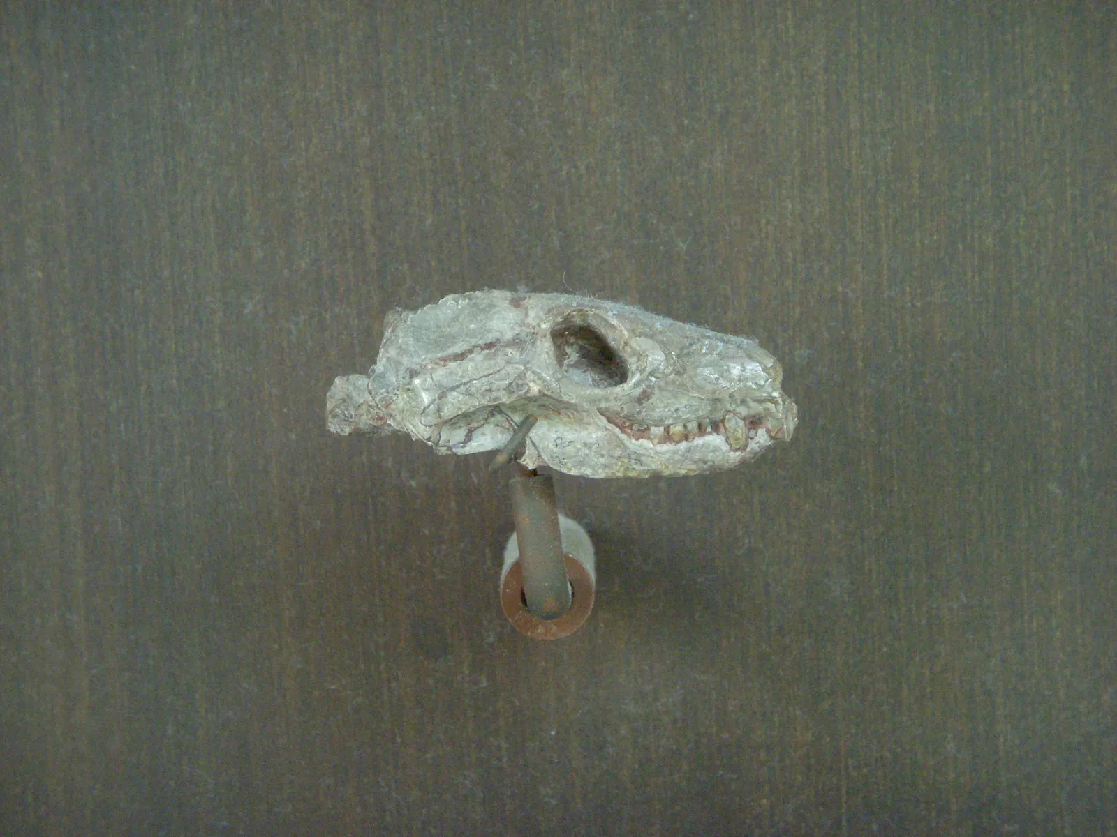
A skull of Thrinaxodon liorhinus, specimen AMNH 5630, on a mount in the American Museum of Natural History, seen from the side. A long row of teeth runs along the lower edge of the skull.
"Thrinaxodon liorhinus skull, AMNH 5630" by Smokeybjb — https://commons.wikimedia.org/wiki/File:Thrinaxodon_liorhinus_AMNH_5630.jpg — CC BY-SA 3.0 (https://creativecommons.org/licenses/by-sa/3.0/)
Same bones, elsewhere:
- Homologous structures: the same matching of bones, followed across a whole forelimb.
- Whale evolution: another change read from fossil bones, this time in the ankles of early whales.
Sources
- Abigail S. Tucker, "Major evolutionary transitions and innovations: the tympanic middle ear", Philosophical Transactions of the Royal Society B 372, no. 1713 (2017): 20150483. doi.org/10.1098/rstb.2015.0483Read: full textText licence: CC BY 4.0 (terms)
- Henry Gray, Anatomy of the Human Body, 20th edition, edited by Warren H. Lewis (Philadelphia: Lea & Febiger, 1918), pp. 219, 221, 227, 230, 266–267, 277 and 1044. archive.org/details/anatomyofhumanbo1918grayRead: pages 219, 221, 227, 230, 266, 267, 277 and 1044, checked against the page images
- Wolfgang Maier and Irina Ruf, "Evolution of the mammalian middle ear: a historical review", Journal of Anatomy 228, no. 2 (2016): 270–283. doi.org/10.1111/joa.12379Read: full text
- Aitor Navarro-Díaz, Borja Esteve-Altava and Diego Rasskin-Gutman, "Disconnecting bones within the jaw-otic network modules underlies mammalian middle ear evolution", Journal of Anatomy 235, no. 1 (2019): 15–33. doi.org/10.1111/joa.12992Read: full text
Cite this page
Feathers, Skeletons & Nests. “Middle Ear Evolution: Two Jaw Bones Became Ear Bones.” first published 17 November 2026. https://feathersskeletonsandnests.com/skeletons/middle-ear-evolution/ (accessed on the date you read it).
How facts are sourcedAll correctionsReport an error on this page (contact@feathersskeletonsandnests.com)
More from the Skeletons drawer
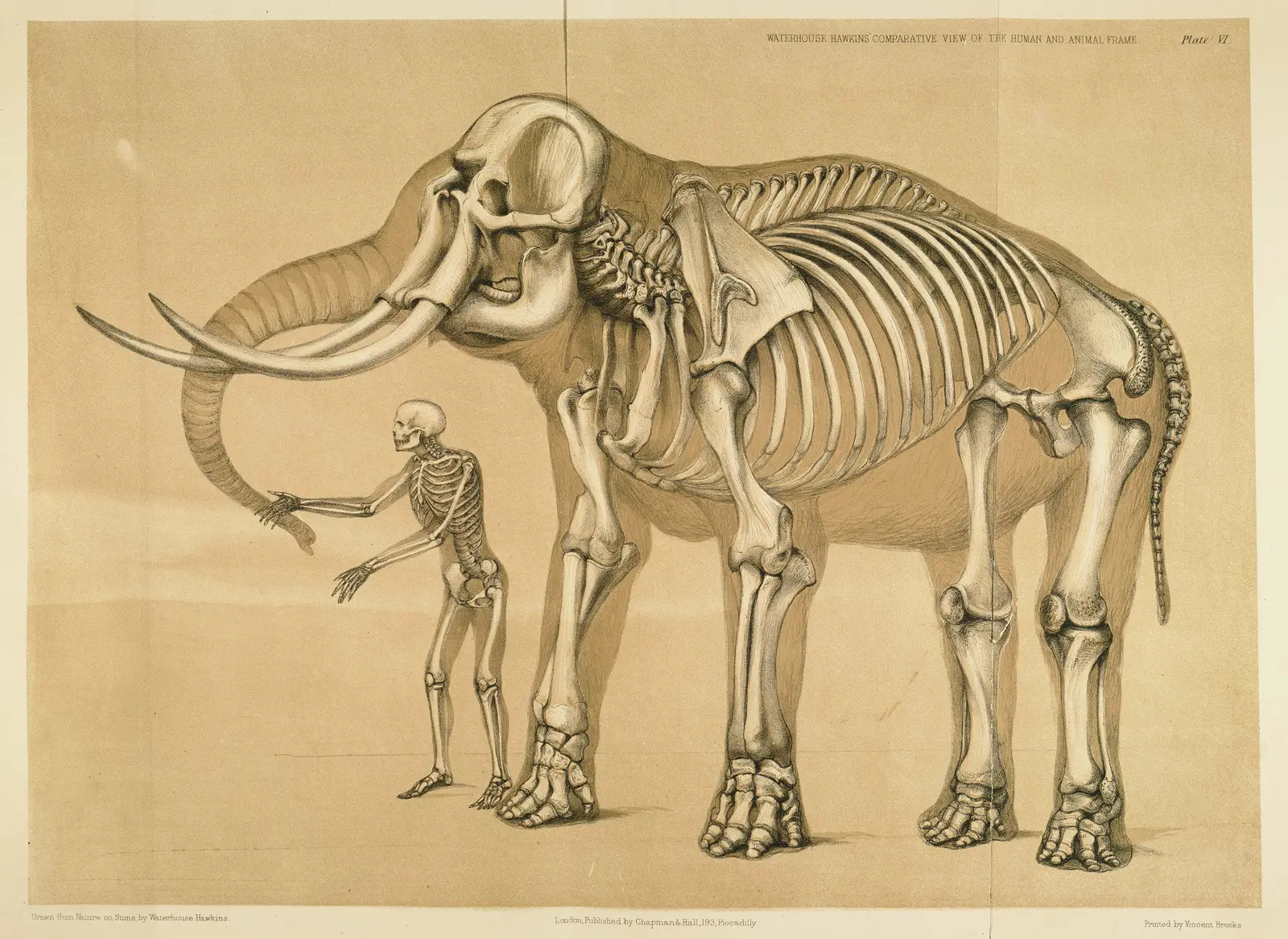
Homologous Structures: Same Bones, Different Counts
- Kind
- In-depth explainer
- Added
Picture credit
"Plate VI, Man and the elephant, from A Comparative View of the Human and Animal Frame" by Benjamin Waterhouse Hawkins — https://commons.wikimedia.org/wiki/File:Comparative_view_of_the_human_and_elephant_frame,_Benjamin_Waterhouse_Hawkins,_1860.jpg — Public domain (US: published 1860; EU: artist Benjamin Waterhouse Hawkins d. 1894, who drew the plate on stone himself (printer Vincent Brooks, dates not checked))
Vestigial Structures: A Whale’s Hip Bones Anchor Muscles
- Kind
- Comparison and reading
- Added
Picture credit
"Skeleton of a baleen whale, from Meyers Konversations-Lexikon, 4th edition" by Unknown author, Meyers Konversations-Lexikon (1888) — https://commons.wikimedia.org/wiki/File:Whale_skeleton.png — Public domain (US: published 1888, before 1931; EU: anonymous work published 1888, more than 70 years ago)
How Many Bones in a Giraffe Neck? Seven, Like Most Mammals
- Kind
- Specimen entry
- Added
Picture credit
"Giraffa camelopardalis (Giraf), skelet, from the Robert Jacob Gordon drawings" by Robert Jacob Gordon / Rijksmuseum — https://commons.wikimedia.org/wiki/File:Giraffa_camelopardalis_(Giraf),_skelet,_RP-T-1914-17-152.jpg — CC0 1.0 (https://creativecommons.org/publicdomain/zero/1.0/)